Leg Bone Diagram / Anatomy Of The Left Lower Leg Doctor Stock. The bones of the leg and foot form part of the appendicular skeleton that supports the many muscles of the lower limbs. The tibia and fibula are two long bones that run parallel to each other, forming the scaffold of the leg and providing attachment points for many muscles. The tibia, commonly known as the 'shin bone', is the largest and most medial of the two.you can palpate its anterior border when you run your finger down the anterior aspect of your leg. In this image, you will find horse leg bone anatomy, femur, stifle joint, tibia, hock joint, splint bone, cannon bone, sesamoid bone, large pastern, small pastern, navicular bone, coffin bone in it. No horse is conformed perfectly.
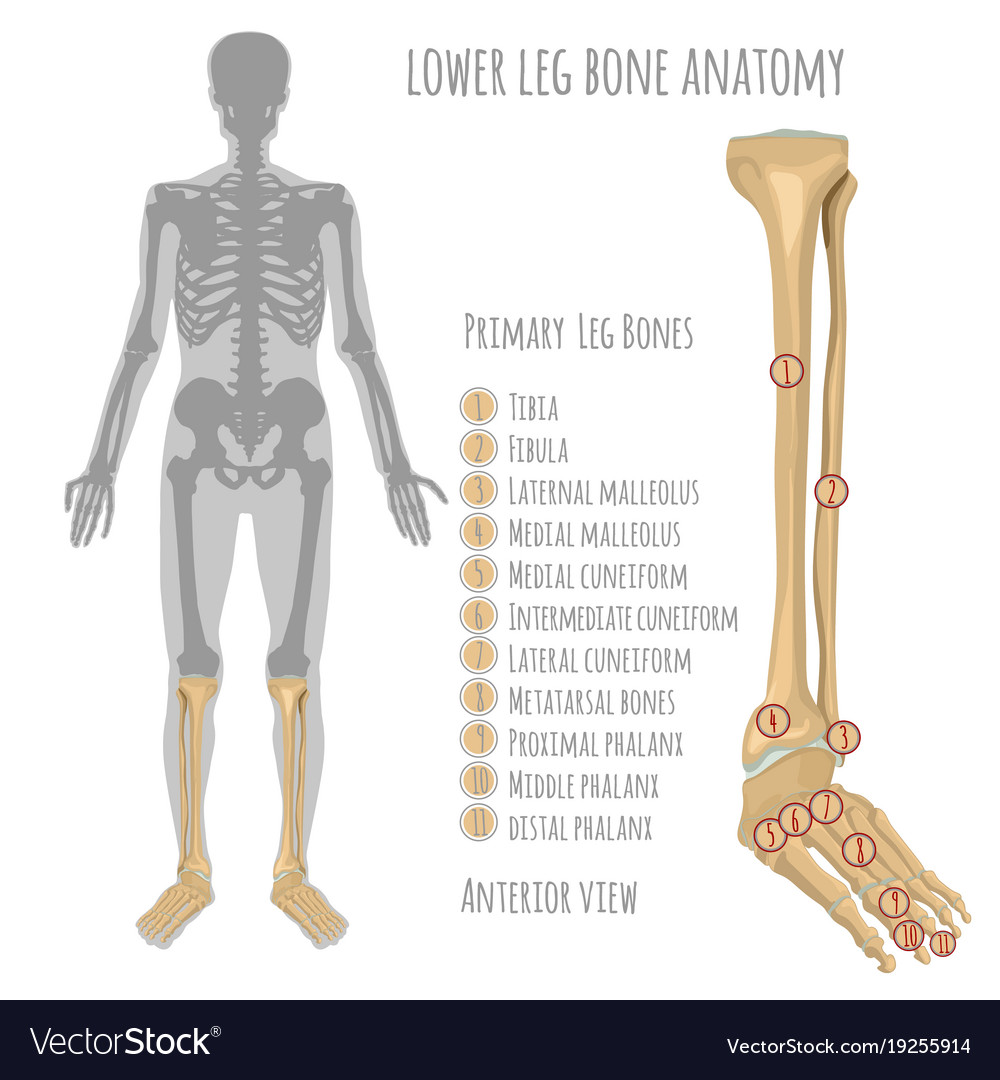
The tibia (also called the shinbone) is located near the midline of the leg. The bones of the leg are the femur tibia fibula and patellathe foot bones shown in this diagram are the talus navicular cuneiform cuboid metatarsals and calcaneus. The patella (kneecap) is the sesamoid bone in front of the knee. The bones of the hip include the femur, the ilium, the ischium, and the pubis. This diagram of a feline skeleton shows you where all of your cat's bones are.
17 214 Leg Bone Stock Photos Pictures Royalty Free Images Istock from media.istockphoto.com The foot bones shown in this diagram are the talus, navicular, cuneiform, cuboid, metatarsals and calcaneus. Diagramme schnell und einfach erstellen. It also separates muscles on the anterior and posterior parts of the leg. Blank leg bones diagram : The knee joint is the largest joint in the body and is primarily a hinge joint, although some sliding and rotation occur. (note, the radius and ulna bones also have this membrane.) this membrane keeps the tibia and fibula together and provides strength and stability for them. The deers skeleton provides protection for the deer it also provides the deer's shape andmovement.the metatarsal bones in a whitetail deer are the longest bones in the deers skeleton. Bone on side of the foot
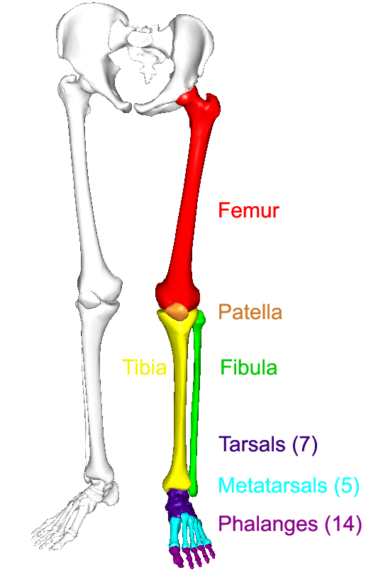
Its lower end helps create the knee joint.
Long bones, short bones, flat bones, and irregular bones.) long bones are longer than they are wide, with spongy bones at both ends and a cavity filled with bone marrow in the shaft. The foot bones shown in this diagram are the talus, navicular, cuneiform, cuboid, metatarsals and calcaneus. The largest and most medial leg bone, forming both the knee and ankle joints. This is a detailed diagram of a horse's hoof. The bones together make up the hip. 6 10 2 votes muscle of the human leg diagram. The following 29 files are in this category, out of 29 total. The hip joint is the uppermost part of the leg where the head of the thigh bone (femur) fits into the socket of the pelvis. The femur is known as a long bone. Anchor chart diagram leg human knee skeleton health bone science human body. Human knee anatomy diagram free vector. Related posts of diagram of leg bones bone of pelvis pics. The bones of the leg are the femur tibia fibula and patellathe foot bones shown in this diagram are the talus navicular cuneiform cuboid metatarsals and calcaneus.
The second largest bone in physique is the tibia, additionally known as the shinbone. The bones of the hip include the femur, the ilium, the ischium, and the pubis. Human knee anatomy diagram free vector. Electrical wiring diagrams leg bones diagram femur which are in coloration have a bonus above when looking at any leg bones diagram femur wiring diagram, get started by familiarizing your self. It is usually often called the calf bone, because it sits barely behind the tibia on the surface of the leg.
Lower Leg Bone Anatomy Royalty Free Vector Image from cdn5.vectorstock.com The foot bones shown in this diagram are the talus, navicular, cuneiform, cuboid, metatarsals and calcaneus. Imagespace skull diagram without labels gmispace com. Knee leg bone diagram clinical practice guidelines : The pubis, ischium, and ilium together constitute the pelvis while the thigh bone is the femur. The foot bones shown in this diagram are the talus, navicular, cuneiform, cuboid, metatarsals and calcaneus leg bone diagram. It also separates muscles on the anterior and posterior parts of the leg. The deers skeleton provides protection for the deer it also provides the deer's shape andmovement.the metatarsal bones in a whitetail deer are the longest bones in the deers skeleton. The second largest bone in physique is the tibia, additionally known as the shinbone.
He leg's main function in the human is for locomotion and support of the rest of the body.
The hip joint is the uppermost part of the leg where the head of the thigh bone (femur) fits into the socket of the pelvis. Learn vocabulary, terms and more with flashcards, games and other study tools. The bones of the leg are the femur, tibia, fibula and patella.the foot bones shown in this diagram are the talus, navicular, cuneiform, cuboid, metatarsals and calcaneus. This diagram of a feline skeleton shows you where all of your cat's bones are. Leg pain can also be caused by blood clots, varicose veins or poor circulation. The bones of the leg and foot form part of the appendicular skeleton that supports the many muscles of the lower limbs. The bones together make up the hip. The patella (kneecap) is the sesamoid bone in front of the knee. The lower leg contains two major long bones, the tibia and the fibula, which are both very strong skeletal structures. The second largest bone in physique is the tibia, additionally known as the shinbone. The largest and most medial leg bone, forming both the knee and ankle joints. The foot bones shown in this diagram are the talus, navicular, cuneiform, cuboid, metatarsals and calcaneus. These bones make up the deers lower leg.
This diagram of a feline skeleton shows you where all of your cat's bones are. In this image, you will find horse leg bone anatomy, femur, stifle joint, tibia, hock joint, splint bone, cannon bone, sesamoid bone, large pastern, small pastern, navicular bone, coffin bone in it. Related posts of diagram of leg bones bone of pelvis pics. The foot bones shown in this diagram are the talus, navicular, cuneiform, cuboid, metatarsals and calcaneus. It is the largest bone in the body and is the only bone in the upper leg.
The Lower Limbs Human Anatomy And Physiology Lab Bsb 141 from s3-us-west-2.amazonaws.com Imagespace skull diagram without labels gmispace com. Leg bones labeled (page 1). Click now to learn more about the bones leg and knee anatomy: The foot bones shown in this diagram are the talus, navicular, cuneiform, cuboid, metatarsals and calcaneus. Simple diagram of leg muscles. The foot bones shown in this diagram are the talus, navicular, cuneiform, cuboid, metatarsals and calcaneus. Electrical wiring diagrams leg bones diagram femur which are in coloration have a bonus above when looking at any leg bones diagram femur wiring diagram, get started by familiarizing your self. At the same time, the bones and joints of the leg and foot must be strong enough to support the body's weight while remaining.
The hip joint is the uppermost part of the leg where the head of the thigh bone (femur) fits into the socket of the pelvis.
Long bones, especially the femur and tibia, are subjected to most of the load during daily activities and they are crucial for skeletal mobility. The bones of the leg and foot form part of the appendicular skeleton that supports the many muscles of the lower limbs. The femur or the thigh bone is closest to the body. Now let's look at the tibia bone, which is the larger of the two leg bones, located medially. He leg's main function in the human is for locomotion and support of the rest of the body. Leg pain can also be caused by blood clots, varicose veins or poor circulation. The bones of the hip include the femur, the ilium, the ischium, and the pubis. The lower leg is comprised of two bones, the tibia and the smaller fibula. The foot bones shown in this diagram one of the beloved filipino beef cuts for is the bulalo, the leg bone section of a cow that is meaty, fatty and full of collagen, not to mention that buttery. Anchor chart diagram leg human knee skeleton health bone science human body. These muscles work together to produce movements such as standing, walking, running, and jumping. The foot bones shown in this diagram are the talus, navicular, cuneiform, cuboid, metatarsals and calcaneus. The hip itself is a ball and socket joint, much like the shoulder.the structures necessary to create this joint are the socket, the joint capsule, muscle, ligaments, and the neck.